Best Bronchoscopy Treatment and Bronchoscopy Doctor in Navi Mumbai
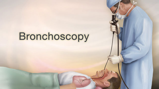
Bronchoscopy is a procedure that allows a pulmonologist in Navi Mumbai to look directly inside the airways and lungs using a thin, flexible tube called a bronchoscope. The bronchoscope is passed through the nose or mouth, past the vocal cords, into the trachea, and down into the bronchi. A small camera and light at the tip of the tube sends a live picture to a screen, allowing the doctor to see the airway lining, spot abnormalities, collect tissue samples, or perform targeted treatments in the same sitting.
Many lung conditions do not give a clear answer on a chest X-ray or CT scan alone. Bronchoscopy closes that gap by giving the chest specialist in Navi Mumbai a direct, real-time view inside the airways, along with the ability to take samples from the exact location where the problem is seen. It is used in diagnosing lung cancer, confirming tuberculosis when sputum tests are negative, evaluating interstitial lung disease, finding the source of bleeding in the airways, and assessing persistent or unexplained respiratory symptoms.
Dr. Vinod B. Chavhan performs bronchoscopy in Navi Mumbai at Chavhan Chest Clinic, Kharghar and Chavhan Chest Clinic, Panvel. He holds an MBBS and FCCP (Pulmonary Medicine, USA) with 17 years of experience in respiratory medicine and has completed over 500 flexible bronchoscopies. He also performs advanced procedures including EBUS-TBNA for patients from Mumbai who need an experienced pulmonologist in Mumbai for airway diagnosis or intervention.
Types of Bronchoscopy
Flexible Bronchoscopy
Flexible bronchoscopy is the most commonly performed type. The bronchoscope is made of fiberoptic material and bends to follow the natural curves of the airway. It is performed under local anaesthesia applied to the throat and nasal passage, with intravenous sedation to keep the patient comfortable. The patient remains conscious but relaxed throughout.
Flexible bronchoscopy in Navi Mumbai is used for:
- Diagnostic sampling: biopsy, BAL, brushings, and TBNA
- Evaluation of haemoptysis: identifying the source of bleeding in the airway
- Assessment of airway abnormalities seen on CT scan
- Clearance of mucus plugs causing lobar collapse
- Foreign body removal from smaller airways
Rigid Bronchoscopy
Rigid bronchoscopy uses a straight, hollow metal tube and requires general anaesthesia. It gives the bronchoscopist direct access to the trachea and main bronchi with better suction capability and allows the use of larger instruments.
Rigid bronchoscopy is used for:
- Removal of large foreign bodies from the central airways
- Management of significant haemoptysis
- Airway stent placement for tumour-related obstruction
- Laser or electrocautery debulking of endobronchial tumours
- Cryotherapy for airway lesions
When Is Bronchoscopy Recommended?
Dr. Chavhan, a chest specialist in Navi Mumbai, recommends bronchoscopy in the following clinical situations:
Diagnostic Indications
Unexplained Haemoptysis (Coughing Up Blood) When a patient coughs up blood without a clear cause on imaging, bronchoscopy is performed to directly identify the site of bleeding within the airway. Endobronchial lesions, inflamed bronchial mucosa, and vascular abnormalities can all be identified.
Lung Cancer Diagnosis and Staging When a mass or lesion is seen on CT scan and lung cancer is suspected, bronchoscopy allows direct biopsy of visible endobronchial lesions or TBNA of involved lymph nodes. EBUS-TBNA improves diagnostic accuracy by using real-time ultrasound to locate and sample mediastinal lymph nodes with a reported sensitivity of 89 to 93% for nodal staging, avoiding the need for surgical mediastinoscopy in most cases.
Tuberculosis: Sputum Smear Negative Cases In patients with a clinical and radiological picture consistent with pulmonary tuberculosis but repeated sputum smear-negative results, bronchoscopy with BAL provides samples directly from the affected lung segment. BAL culture positivity in sputum smear-negative TB can reach up to 90%, significantly improving diagnostic yield over repeated sputum testing alone.
Interstitial Lung Disease (ILD) ILD includes conditions such as hypersensitivity pneumonitis, sarcoidosis, pulmonary fibrosis, and connective tissue disease-related lung involvement. BAL fluid analysis, including macrophage, lymphocyte, neutrophil, and eosinophil differential counts, provides information that helps narrow the ILD diagnosis when combined with HRCT patterns and clinical history. EBUS-TBNA is additionally useful in sarcoidosis where mediastinal lymph node enlargement is present.
Persistent or Recurrent Pneumonia When pneumonia recurs in the same lobe or segment, or fails to clear with antibiotics, bronchoscopy evaluates for an underlying obstructing lesion, endobronchial abnormality, or an unusual organism not captured on standard sputum culture.
Abnormal Chest Imaging A shadow, consolidation, nodule, or mass seen on chest X-ray or CT that remains unexplained after initial investigation is a standard indication for bronchoscopy to obtain tissue or fluid samples for pathological or microbiological analysis.
Mediastinal or Hilar Lymphadenopathy Enlarged lymph nodes in the mediastinum or lung hilum, seen in sarcoidosis, lymphoma, tuberculosis, or lung cancer, can be sampled using EBUS-TBNA without requiring a surgical procedure.
Chronic Unexplained Cough In patients with a persistent chronic cough lasting more than 8 weeks where common causes have been excluded, bronchoscopy evaluates for endobronchial lesions, tracheobronchomalacia, or inflammatory airway changes.
Stridor and Airway Narrowing An abnormal breathing sound caused by turbulent airflow through a narrowed airway is directly assessed with bronchoscopy to identify its level and cause, whether from a tumour, stricture, vocal cord abnormality, or tracheal compression.
Therapeutic Indications
- Removal of foreign bodies from the airway
- Clearance of mucus plugs causing lobar collapse
- Balloon dilation of benign airway strictures
- Endobronchial stenting for malignant central airway obstruction
- Electrocautery or cryotherapy for endobronchial tumours
- Control of airway bleeding with adrenaline instillation or cautery
Bronchoscopic Procedures Explained
Bronchoalveolar Lavage (BAL)
BAL is performed by wedging the bronchoscope tip into a segmental or subsegmental airway and instilling sterile normal saline, typically in aliquots totalling 100 to 200 ml, which is then suctioned back out. The returned fluid carries cells, organisms, and inflammatory mediators from the alveolar level of the lung.
BAL fluid is sent for:
- Differential cell count (macrophage, lymphocyte, neutrophil, eosinophil proportions)
- Microbiology: bacterial, fungal, and mycobacterial cultures
- Cytology for malignant cells
- Specific staining for AFB (acid-fast bacilli) and Pneumocystis jirovecii
BAL is particularly valuable in diagnosing infections in immunocompromised patients, evaluating ILD, and confirming sputum smear-negative tuberculosis.
Endobronchial Biopsy
When an abnormal area of bronchial mucosa is seen (a growth, ulceration, nodule, or irregular vascular pattern), biopsy forceps are passed through the bronchoscope to take a small tissue sample. This sample is sent for histopathological examination to confirm or exclude malignancy, granulomatous disease, or infection.
Transbronchial Lung Biopsy (TBLB)
In TBLB, biopsy forceps are advanced beyond the visible airways into the peripheral lung tissue under fluoroscopy guidance. This technique obtains lung parenchymal tissue without surgery. It is used in ILD, suspected lung transplant rejection, and peripheral lung infiltrates.
EBUS-TBNA (Endobronchial Ultrasound)
EBUS adds a miniaturized ultrasound transducer to the tip of the bronchoscope. This allows real-time imaging of structures outside the airway wall, including mediastinal and hilar lymph nodes, vascular structures, and parabronchial masses. A fine needle is then advanced through the bronchoscope wall under direct ultrasound guidance to aspirate cellular material from these structures.
EBUS-TBNA is used for staging of lung cancer, diagnosis of sarcoidosis with mediastinal lymph node involvement, and sampling of unexplained mediastinal or hilar lymphadenopathy. Dr. Chavhan performs EBUS-TBNA at Chavhan Chest Clinic. Full details are on the EBUS-TBNA page.
Bronchial Brushings and Washings
Cytology brushes are passed through the bronchoscope to scrape cells from visible lesions or peripheral areas of the lung. Bronchial washings involve instilling and aspirating small amounts of saline from a localized area. Both provide cytological material that can detect malignant or pre-malignant cells.
Pre-Procedure Preparation
Before your bronchoscopy at Chavhan Chest Clinic, Kharghar or Chavhan Chest Clinic, Panvel, the following steps are required:
Fasting You must have nothing to eat or drink for at least 6 hours before the procedure. This reduces the risk of aspiration if you cough or gag during the procedure.
Medication Review Inform Dr. Chavhan’s team of all medications you take. Blood thinners including warfarin, clopidogrel, and aspirin need to be paused before the procedure at a schedule decided by the doctor, as they increase the risk of bleeding if a biopsy is taken. Diabetes medications, particularly insulin and oral hypoglycaemics, need adjustment on the day of fasting. Do not stop any medication without specific instruction from the team.
Pre-Procedure Assessment Your oxygen levels, blood pressure, heart rate, and recent blood reports including coagulation tests and platelet count will be reviewed before the procedure. A recent chest CT or X-ray report helps Dr. Chavhan plan which airways and areas to evaluate.
Companion Required Bring a family member or companion who can take you home after the procedure. The sedative used during bronchoscopy reduces alertness for several hours. You should not drive or operate machinery on the day of the procedure.
What Happens During Bronchoscopy
On arrival, you will be connected to a pulse oximeter to monitor oxygen saturation, along with ECG leads for heart rate monitoring and a blood pressure cuff. These remain in place throughout.
Local anaesthetic spray (lignocaine) is applied to the back of the throat and nasal passage to suppress the gag reflex and reduce discomfort. Intravenous sedation is given through a small cannula in your hand or arm. You will feel drowsy and relaxed within a few minutes.
The flexible bronchoscope is then passed through the nose or mouth. The vocal cords are inspected and additional anaesthetic is sprayed onto them before the scope passes into the trachea. Dr. Chavhan systematically examines the trachea, carina, main bronchi on both sides, and the segmental and subsegmental airways as far as required.
Depending on what is found and what was planned, one or more of the following are performed: BAL, biopsy, brushings, washings, or EBUS-TBNA. Oxygen is delivered via nasal prongs throughout to maintain adequate oxygen saturation.
The procedure takes 20 to 45 minutes for routine diagnostic bronchoscopy.
Recovery After Bronchoscopy
After bronchoscopy, you will be monitored in a recovery area. Oxygen saturation, heart rate, and blood pressure are checked until the sedative effects have resolved and you are fully awake. This typically takes 60 to 90 minutes.
Common after-effects that resolve on their own:
- Sore throat or hoarse voice. This settles within 1 to 2 days
- Mild cough. This clears as the airway recovers
- Drowsiness from the sedative. This lasts a few hours
- Blood-streaked mucus for 24 to 48 hours if a biopsy was taken. This is expected
Instructions for the day of the procedure:
- Do not eat or drink until the numbness in your throat has fully resolved, usually 2 to 3 hours after the procedure
- Do not drive or make important decisions that day
- Resume regular medications as advised by the team
Most patients return to normal daily activities the following day.
Risks and Complications
Bronchoscopy is a safe procedure. Serious complications occur in fewer than 1% of cases.
Minor and common:
- Sore throat and mild cough: very common and temporary
- Low-grade fever in the 24 hours after BAL. This settles on its own
- Small amounts of blood in sputum after biopsy. This resolves within 1 to 2 days
- Transient drop in oxygen saturation during the procedure. This is managed with supplemental oxygen
Less common:
- Significant bleeding at the biopsy site. This occurs in approximately 1 in 30 patients who have a lung biopsy and is managed with medications or direct cautery in most cases
- Bronchospasm: airway tightening triggered by the procedure, managed with bronchodilator treatment
Rare:
- Pneumothorax (collapsed lung). This occurs in approximately 1 in 50 patients after transbronchial lung biopsy. Most resolve with oxygen and a small number require a chest drain
- Laryngospasm or vocal cord spasm
- Cardiac arrhythmia: more likely in patients with pre-existing heart disease. ECG monitoring during the procedure allows early detection
Dr. Chavhan’s team monitors oxygen saturation, heart rate, blood pressure, and ECG continuously during every bronchoscopy at Chavhan Chest Clinic, Kharghar and Panvel.
Contraindications
Bronchoscopy may not be suitable in all patients. Relative contraindications include:
- Severe uncorrected coagulation disorder or very low platelet count, especially if biopsy is planned
- Unstable cardiac arrhythmia or recent heart attack
- Severely reduced lung function with resting oxygen saturation below 90% on air that cannot be corrected with supplemental oxygen
- Uncontrolled severe bronchospasm
In some of these cases, the procedure may still be performed if the clinical need outweighs the risk, with additional precautions in place.
Results and Follow-Up
If a biopsy or BAL sample was taken, the specimen is sent to a pathology and microbiology laboratory. Histopathology results are typically available within 5 to 7 days. Mycobacterial cultures for TB confirmation take 4 to 8 weeks. Cytology results are usually available within 3 to 5 days.
Dr. Chavhan’s team will contact you when the report is ready and schedule a follow-up appointment to discuss the findings and the next steps in your treatment.
Related Services at Chavhan Chest Clinic
EBUS-TBNA (Endobronchial Ultrasound) is performed alongside bronchoscopy when mediastinal or hilar lymph nodes need to be sampled. An ultrasound probe attached to the bronchoscope allows Dr. Chavhan to see and needle lymph nodes outside the airway wall in real time. This is the standard technique for staging lung cancer and diagnosing sarcoidosis without open surgery.
Lung Cancer Diagnosis and Management at Chavhan Chest Clinic covers the full diagnostic pathway from CT imaging review through bronchoscopic biopsy and EBUS-TBNA tissue confirmation. Bronchoscopy is often the procedure through which a lung cancer diagnosis is first established, making it a central step in the oncology workup Dr. Chavhan coordinates for his patients.
Tuberculosis Diagnosis and Treatment in sputum smear-negative patients frequently requires bronchoscopy to collect BAL samples directly from the affected lung segment. Dr. Chavhan has extensive experience managing pulmonary tuberculosis, including cases where standard sputum testing has not provided a definitive result and bronchoscopy is needed to confirm the diagnosis.
Interstitial Lung Disease (ILD) evaluation often involves bronchoscopy with BAL fluid analysis and, in selected cases, transbronchial lung biopsy. Dr. Chavhan assesses patients with ILD including hypersensitivity pneumonitis, sarcoidosis, and pulmonary fibrosis, using bronchoscopic findings alongside HRCT imaging and clinical history to guide diagnosis and treatment planning.
Clinic Locations
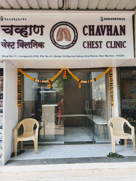
Chavhan Chest Clinic - Panvel
📍 Shop No-21, Indrsprasht CHS, Plot No-1, Sector-10, Khanda Colony, New Panvel East, Navi Mumbai, Maharashtra 410206
Phone: +91 9004985020
Get Location
Chavhan Chest Clinic - Kharghar
📍 Shop No 312-313, 3rd Floor, Pacific Building, Shilp Chowk, Sector 13, Kharghar, Navi Mumbai, Maharashtra 410210
Phone: +91 9820676822
Get Location
Kokilaben Dhirubhai Ambani Hospital
📍 Thane-Belapur Road, Kopar Khairane, Navi Mumbai, Maharashtra 400710
Phone: +91 9004985020
Get Location
Sanjivani Speciality Clinic - Vashi
📍 Office No. 706, JK Chambers Building, Sector 17, Vashi, Navi Mumbai, Maharashtra 400703
Phone: +91 9820676822
Get Location